GOLGI BODIES:
Golgi complex or Golgi bodies or Golgi apparatus or simply Golgi is a cellular organelle present in most of the cells of eukaryotic organisms. It is a cup-shaped organelle that is located near the nucleus. It packages proteins into membrane-bound vesicles inside the cell before they are sent to their destination. This Golgi body was first discovered by an Italian biologist "Camello Golgi" in 1898. It's structure can be explained below:

Structure of the Golgi body:
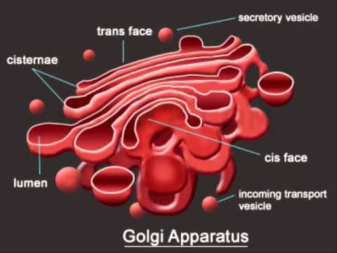
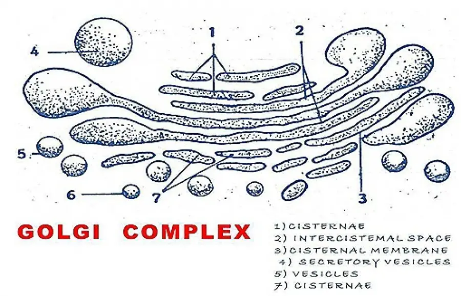
Under the electron microscope, the Golgi body is seen to be composed of stacks of membranous flattened sacs and vesicles concerned with the cell secretion. The Golgi apparatus is morphologically similar in both plant and animal cells. However, it is extremely pleiomorphic. In some cell types, it's shape and form may vary depending on cell type. Numerous cisternae are associated with each other and appear in a stock like aggregation and a group of these cisternae is called "dictyosomes". This group of dictyosomes makes up the cells of the Golgi body.
The size of the Golgi body is variable. It is larger and well developed in active cells like gland cells and nerve cells whereas poorly developed in muscle cells. The position of Golgi is relatively fixed for each cell type. It usually occupies near the nucleus. There is a clear region around the Golgi body. It has no ribosomes, mitochondria, chloroplast, and even endoplasmic reticulum which is called as "zone of exclusion". Typically, Golgi apparatus appears as a complex array of the interconnecting tubules, vesicles, and cisternae.
Cisternae:
- The cisternae are elongated flattened sacs of Golgi complex filled with fluids and piled one upon the other to form a stack.
- They are arranged in a parallel bundle one above the other.
- In most of the cells, the number of cisternae varies from 3 to 7 in a stack. Other cell types may have as many as 10-20 cisternae.
- Each stock of cisternae forms a dictyosome which may contain 56 golgi cisternae in an animal cell, 20 or more cisternae in plant cells.
- Each cisterna is bounded by a smooth unit membrane having a lumen varying from a range 500-1000nm.
- The adjacent cisternae are cemented together by a cementing substance called intracisternal material.
- In certain cases, the cisternae contain pores and they are said to be "fenestrated golgi".
- The cisternae are slightly curved and the cisternae have convex and concave surfaces, due to which the golgi complex has two sides, namely forming face and maturing face.
- The convex surface is the forming face or cis face. This face is towards the nucleus or ER. Here new lamellae are added from the endoplasmic reticulum.
- The concave surface is the maturing face or trans face. The transface is towards the plasma membrane. Here large secretory vesicles are budded off.
- Thus the cisternae are continually receiving the lamellae on the forming face and losing membranes on the maturing face through the formation secretory vesicles.

Tubules:
- A complex array of associated vesicles and anastomosing tubules surround the dictyosome and radiate from it. In fact, the area of dictyosome is a fenestrated lace-like structure.
Vesicles:
- These are small droplet like the structure of about 40 (Angstroms) in diameter.
- They are closely associated with the periphery of the cisternae.
- They develop either by budding or by the constriction of the ends of the cisternae.
Functions of Golgi bodies:

- This helps in the formation of the cell wall and the plasma membrane.
- The golgi bodies help in the biogenesis of lysosomes where primary and secondary lysosomes are involved.
- Golgi complex carries out a trafficking function in the transport of biosynthetic products. They are transported within the cells (intracellular) or to outside the cells (intercellular transport).
- The Golgi complex helps in the production of proteoglycans which are molecules present in the extracellular matrix.
- The golgi body in the animal cells involves the packaging of certain enzymes such as mucus, lactoprotein, etc.
- They are also involved in the transport of lipid molecules around the cell.
- It is also the major site of synthesis of carbohydrates.

- Courtesy by google images
Very reasonable
ReplyDeleteSimply super
ReplyDeleteEasy to understand
ReplyDeleteSuperb bioreaders
ReplyDelete