Search here

Showing posts with label Cell biology. Show all posts

Wednesday, 6 January 2021

 Vacuoles:-

                              Vacuoles are the membrane-bound organelles found in all plant cells, bacterial, fungal, and animal cells too. It is a storage structure in a cell. These are the empty space organelles found in the cytoplasm and filled with watery fluid that contains various substances. These vacuoles are the important cell organelles in the cell as they help in the storage of nutrients required by a cell to survive and can also store the waste from the cell thereby preventing the cell from contamination. Plant cells store nutrients, metabolites, and waste in their vacuoles and also use them for transporting from one cell to another. The vacuoles in plants are larger than the animal cells.


Structure of vacuole:-

             It is a membrane-bound structure present in the cellular matrix of a cell. Generally, there is no basic shape or size for this vacuole. The vacuoles are usually small during immature or undividing stages and arise initially to become a large ones. Actually plant cells have larger vacuoles than animal cells as they require more water, organic and inorganic components for the proper functioning of cells.  A vacuole is surrounded by a membrane called "tonoplast" or "vacuolar membrane". 

                      This tonoplast separates the vacuolar contents from the cell's cytoplasm. This membrane mainly involves in the regulation of ions in the cell. It also helps in isolating the particles that are thought to be a threat to the cell. The components of the vacuole is known as 'cell sap' that differs entirely different from the cytoplasm. There may be several vacuoles in a cell and this tonoplast helps to separate from the cytoplasm. These vacuoles are relatively similar to lysosomes as they also contain a wide range of hydrolytic enzymes.


Types of Vacuoles:- 

                  There are various types of vacuoles present in the cells and some of them are:-

(i)Sap vacuoles:- These sap vacuoles are found mostly in plant cells and consist of a number of transport systems for the passage of different substances. A large central vacuole is present in higher plants. The fluid present in this vacuole is known as 'sap'. 


(ii)Contractile vacuoles:- These are found in freshwater algal cells and some protists such as paramoecium. These contractile vacuoles take part in osmoregulation and excretion.

(iii)Food vacuoles:- It is formed by the fusion of lysosome and phagosome and it consists of digestive enzymes by which food and the other nutrients are digested. These food vacuoles are found in protists, protozoa, and other higher animals, etc.

(iv)Gas vacuoles:- These are also called as 'air vacuoles' and store gases. Apart from storing gases, they also help in providing buoyancy, mechanical strength, and protection from harmful radiations. These vacuoles are found in prokaryotes.

Gas vacuoles

Functions of vacuoles:-

  • Storage:- Vacuoles help in the storage of salts, minerals, organic acids, and other proteins within the cell. A large number of lipids are stored in the vacuoles. Some waste products are also stored here.
  • Transportation in plant cells:- Proteins found in the tonoplast control the flow of water in and out of the vacuole through active transport and also pump potassium ions in and out of the vacuolar interior. It also helps in endocytosis and exocytosis of various substances and lysosomes are the vesicles that intake food and digest it.
  • Turgor pressure:- Vacuoles are completely filled with water by which it exerts a force on the cell wall. This is known as turgor pressure. Also, the salts present in vacuole add to the osmotic activity of the vacuole thereby contributing to turgor pressure. This pressure helps in cell elongation, withstand extreme conditions, supporting plants in an upright position, and also provides a shape to the cell.
  • The vacuole pushes up all the contents of the cell's cytoplasm against the cellular membrane thus keeping the chloroplasts closer to the light so that the light-absorbing efficiency is improved.
  •  The pH of the plant vacuoles may be as 9 to 10 due to large quantities of alkaline substances or as low as 3 due to the accumulation of quantities of acids.
  • In fungal cells, vacuoles are involved in many processes such as homeostasis of cell pH, osmoregulation, the concentration of ions and degradative processes, etc.
 

Saturday, 2 January 2021

 Nucleus:-

                                The nucleus is an oval-shaped membrane-bound organelle present in all eukaryotic cells. It is present both in plant and animal cells. It is large and present in the center of a cell. It is a structure that contains the cell's hereditary information and helps in controlling the growth and reproduction of the cell. It is the most integral component of the cell and contains DNA which controls the growth and function of the cell and helps in coordinating the cell activities. The nucleus here is similar to the brain in it's functions and hence known as the "Brain of the cell"

                 It was first discovered by "Robert Brown" in 1831 when he was scrutinizing the epidermis in a collection of orchids with his microscope.  He identified a small opaque spot in the cell and later he noted that this spot can be observed in the early stages of pollen formation. He then further realized that this part is an important component in the cell and named it as "Nucleus". 


Structure of Nucleus:-

                      It is a typically membrane-bound structure and the most prominent organelle in a cell. It is present in all eukaryotic cells except in few cells like mammalian cells etc. The nucleus may be round, oval, or disc-shaped depending on the type of cell. Structurally, the cell nucleus consists of the following parts. They are as follows;-

  1.  Nuclear membrane
  2. Nucleoplasm
  3. Nucleolus
  4. and chromosomes 

1.Nuclear membrane:-

  •  It is a double layered membrane that surrounds the nucleus and protects it from any mechanical injuries.
  •  As it encloses the nucleus, it is also called a "Nuclear envelope". 
  • It helps in the entry and exit of material into the nucleus and also separates the nucleus from the other parts of the cell.
  •  A liquid-filled space is present between the two layers of the membrane called the 'perinuclear space'.
  • The nuclear membrane is perforated with numerous pores called "nuclear pores". 
  • The "Nuclear pores" are the sites for the exchange of large molecules between the nucleus and cytoplasm.

2.Nucleoplasm:-

  •  The liquid-filled matrix present in between the two layers of the membrane is called as 'nucleoplasm'.
  •  It is also called the  'karyoplasm'


3.Nucleolus:-

  • It is a small, spherical-shaped structure found in the nucleus of both plant and animal cells.
  • It plays a vital role in the synthesis of RNA and in the formation of ribosomes.
  • Some of the eukaryotic cells have nucleus consisting of more than 4 nucleoli.
  • The nucleolus disappears when a cell undergoes cell division and again reforms after the cell is formed.

4. Chromosomes:-

  • Chromosomes consist of DNA that contains hereditary information and promotes cell growth, cell development, and reproduction.
  • These are self-reproducing thread-like structures packed with DNA located in the nucleus.
  • When a cell is resting, ie, not dividing the chromosomes are organized into long structures called 'chromatin'.
    (for more details on this topic, refer to the chromosomes and chromatin page in the blog)

Nucleus in plant cell:-

                                  The nucleus in the plant cells can be different in different plants. The various forms of nucleus present in the plant can be given in the following way;-

  • Uninucleate cell: Plant cell which contain only a single nucleus and also referred to as monokaryotic cell
  • Binucleate cell: Plant cell which contains two nuclei at a time and also referred to as dikaryotic cell. For eg; paramoecium
  • Multinucleate cell: Plant cell which contains more than 2 nuclei at a time and also referred as polynucleate cell. For eg; latex cells in plants, bonemarrow cells in animals
  • Enucleate cell: Plant cell without a nucleus. For eg; mature sieve tubes of phloem, RBCs of mature mammals.

Functions of Nucleus:-

  1. The nucleus is responsible for cell division, growth, differentiation, and protein synthesis too.
  2. It helps in the exchange of DNA and RNA between the nucleus and the other parts of the cell.
  3. It is the control centre of the organism as it regulates the integrity of the gene and gene expression.
  4. It controls the hereditary characters of a cell organism.
  5. The nucleus is the site of transcription where mRNA is produced for protein synthesis.
Robert brown

  • Courtesy by google images 


Thursday, 31 December 2020

 Chromatin:-

                                     In eukaryotes, the genetic material ie, DNA is complexed with proteins in a specialized structure called as "Chromatin". This chromatin consists of double-stranded DNA to which large amounts of protein and a small amount of RNA are added. This chromatin usually engages with functions like repair, replication, and recombination, etc.  The dynamic structure of this chromatin influences it to work functionally in the genome. 

Nucleosome:                                     

                  The fundamental unit of chromatin is called as 'Nucleosome'. It comprises of DNA, RNA, and some basic proteins such as histones and non-histones (acidic proteins). The amount of RNA and non-histone proteins is variable depending on different chromatin structures whereas there are fixed proportions of DNA and histone proteins in a 1/1 ratio. The histones that are attached to the DNA act as 'anchors' that help in the winding of the components. The non-histones are very heterogenous as they vary in different tissues and include DNA and RNA polymerases among other enzymes. The nucleosome is composed of two main parts:

  • the core particle and
  • the linker region that adjoins the core particles.

      The core particle is highly conserved and composed of 146 base pairs of DNA wraps around the histone octamer (8 octa core proteins).

       The linker histone links the entry or exit of the DNA strand on the nucleosome.


        Histones:-

                        Histones are the basic proteins present in the DNA of chromatin. There are of five types each one present in large amounts. Histones are small proteins that are basic because of their high content of basic amino acids such as arginine and lysine. Now besides being basic,  they help in binding tightly to DNA, which is an acid. The four main histones are H2A, H2B, H3 and H4. 

             These four histones are present in equimolar units, but H1 is not conserved. It is present only once per 200 basic pairs. It is loosely attached to the chromatin and is not a component of the nucleosome core unit ie, DNA- histone structure. It is bound to the linker segments of DNA that joins the neighbouring nucleosomes. Under the electron microscope, the nucleosome of eukaryotic nuclei it was found that the chromatin had the repeating structure of beads about 10nm of diameter connected by a string. It appeared as "beads-on-string" structure.  


Types of Chromatin:-

     There are mainly two types of Chromatin. They are explained below:

  1. Heterochromatin:-

  •   It is a tightly packed form of chromatin silencing gene transcription ie, the genes or transcription sequences present in them are inactive. 
  • Heterochromatin is usually present in the nucleus towards the periphery,
  • This heterochromatin is difficult to analyze because of it's condensed state and repetitive DNA sequences.
  • It is characterized by intense stains when stained with nuclear stains.
  • It is a structure that does not alter in its condensation throughout the cell cycle and it is much more condensed than the euchromatin.
  • The tightly packaged DNA in the heterochromatin prevents the chromosomes from various protein factors that lead to the binding of DNA.
  • It also helps to prevent the inaccurate destruction of chromosomes by endonucleases.
  • Heterochromatin has various functions such as gene regulation, chromosomes integrity etc,
  • Telomeres, centromeres, bar bodies, genes 1,9,16 of human beings are some examples of heterochromatin.
  • There are mainly two types of heterochromatin. They are as follows:

                     a)Constitutive heterochromatin

                      b)Facultative heterochromatin

Constitutive heterochromatin usually contains and packages the same sequences of DNA in all the same species. It is also repetitive and usually coincides with the structural regions such as telomeres and centromeres. The genes of this constitutive heterochromatin might affect the genes of the tightly-packed ones.                                                                                For eg: In humans, the Y-chromosome in men constitutes this constitutive heterochromatin

Facultative heterochromatin is that region of the chromosome that is heterochromatic in some cells and euchromatin in other cells. It is composed of transcriptionally active genes that adopt the structural and functional characteristics of heterochromatin, but is not as repetitive as the constitutive one.                                                                                               For eg: In humans, one of the X chromosomes in women is inactivated as facultative heterochromatin while the other is expressed as euchromatin. 

2.Euchromatin:-

  • It is less condensed and contains the most actively transcribed genes. It is lightly packed DNA that is characterized by less intense staining.
  • This is present in the interior of the nucleoplasm and the DNA sequences are transcriptionally active.
  • The DNA in euchromatin is unfolded to form a beaded structure, unlike heterochromatin to which histone proteins are folded.
  • In euchromatin, the DNA is lightly bound and the genes are active or will be active during the growth.
  • Euchromatin forms a more significant part of the genome. It makes 90-92% of the genome in the human being.
  • Euchromatin is found in both eukaryotes and prokaryotes. and it exists only in one form ie, constitutive form.
  • There are various other chromosomes other than heterochromatin which are examples of  euchromatin 

Significance of Chromatin:

                                Chromatin is meant for efficiently packaging of DNA into small volumes to fit into the nucleus of a cell and protect the DNA structure and its sequence. Packaging DNA into chromatin allows for cell divisions and prevent chromosome breakage besides controlling gene expression and DNA replication.

  • courtesy by google images

Monday, 28 December 2020


CHROMOSOMES:-

                                  Chromosomes are self-reproducing thread-like structures packed with the DNA located inside the nucleus. These are derived in the form "chromo" which means "color" and "soma" means "body" and are easily stained with dyes. They are the vehicles of heredity ie, concerned with the transmission of characters from generation to generation. Each chromosome is made up of DNA tightly coiled many times around proteins called "histones" that support its structure. 

                    Chromosomes were first observed by "Hofmeister" in 1848 in the nuclei of pollen mother cells of Tradescantia. However, the term chromosome was first named and used in 1888 by "Waldeyer". Chromosomes appear as dark-stained bodies during mitosis when the cells are stained by a suitable basic dye and viewed under a light microscope.


                         The chromosomes are large, linear present in the nucleus. Each chromosome typically has one centromere and one or two arms that project out from the centromere. During mitosis, the chromosomes split longitudinally into two chromatids.

                The two chromatids are attached to the centromere. Each chromosome is divided into three parts namely:

  1. Pellicle: It is the thin outer substance that surrounds the chromosome.
  2.  Matrix: It is the ground part of the chromosome which consists of chromonemata.
  3.  Chromonemata: These are two identical, spirally coiled threads embedded in the matrix of each chromosome.


                           The number of chromosomes varies from species to species. But the number remains constant among the members of the same species. The lowest number of chromosomes is 2 which occurs in Ascaris megalocephala. And the maximum number of chromosomes is 1700 which occurs in radiolarian.

The size of the chromosome ranges from 0.1-30 microns. The diameter varies from 0.2-2 microns. In general, plants have larger chromosomes than animals. For eg; the plant Trillium has chromosomes with the length of 32 microns at the metaphase.  The length of the human chromosome varies from 4-6 microns.

There are some other points to be noted. Generally, the chromosomes are arranged in pairs.

  • A pair of similar chromosomes are called “homologous chromosomes”.
  •  The somatic cells contain two sets of chromosomes which are called diploid numbers and are represented by “2n”.
  • The gametes contain only one set of chromosomes which is called the haploid number and is represented as “n”.
  •   Sometimes a cell may contain more than two sets of chromosomes, and this number is called as “polyploid number (3n,4n,5n)

Types of Chromosomes:-

1. based on the position of centromere:-

                                 The shape of a chromosome is largely determined by the position of its centromere ie, a small structure in chromonemata that divides a chromosome into two arms. The short arm is represented as the ‘p’ arm whereas the long arm is the ‘q’ arm. On this basis, chromosomes are classified into four types. They are the following;

a. Telocentric:- The centromere is located at the terminal end of the chromosome, so the chromosome has just one arm. Such chromosomes are rare and exist normally in certain species of Protozoa

b. Acrocentric:- These are rods like chromosomes having a very small arm and a very long arm. The centromere occupies a subterminal position. This is a characteristic of Locus.

c. Sub-metacentric:- These chromosomes are L-shaped having unequal arms. The centromere is slightly away from the midpoint.

d. Metacentric:- These chromosomes are ‘V’ shaped. They have arms equal in length. Here the centromere lies in the middle of a chromosome. This is a characteristic of Amphibia.


2. based on the number of centromeres:-

                                    The chromosomes are divided into five types depending on the number of centromeres. They are

a. Monocentric : consists of one centromere

b. Dicentric: consists of two centromere

c. Polycentric: consists of more than two centromeres

d. Acentric: It does not consist of any centromere. These are the freshly broken parts of chromosomes that do not survive for long.

e. Diffused or non-located with indistinct centromere diffused throughout the length of the chromosome.

3.   Human Chromosomes:-

                      Humans have 23 pairs of chromosomes (46 Chromosomes) in their cells.  The human chromosomes are of two types. They are:-

a.   Allosomes:- Genetic traits of the sex of a person are passed on to sex chromosomes or allosomes. Humans have one pair of sex chromosomes.

b.   Autosomes:- The rest of the genetic information is present in the remaining 22 pairs of chromosomes known as autosomes.

Functions of Chromosomes:-

  •    Self duplication:- They help in transferring the characters from one generation to another generation or from parents to offsprings.
  •   Chromosomes controls the biological process in the body of an organism.
  • These control the cell metabolism by directing the formation of necessary protein in our body and maintain the order of DNA.
  •  They help in cell differentiation during the development of cell and controls the cell division.
  •    A chromosome also helps in determining the sex of the individual.

  Giant chromosomes:-

                         The giant chromosomes are exceptionally larger ones. These are described as unusual chromosomes by A.M.Winchester.There are two types of giant chromosomes namely

a)   Polytene chromosomes

b)  Lampbrush chromosomes

    a) Polytene chromosomes: 

                These were discovered by Balbiani in 1881. They are found in salivary glands, gut cells, and fat body cells of insects. It is a giant chromosome that is larger in size. For eg; Drosophila melanogaster is 1000 times larger than the somatic chromosomes. The larger size of the chromosome is due to the presence of many longitudinal strands called chromonemata.

        The polytene chromosomes contain two types of transverse bands, namely dark bands and inter bands 

  •   The dark bands contain more DNA and less RNA
  •    The inter bands contain more RNA and less DNA.  

     The bands of polytene chromosomes become enlarged at certain times to form swellings called “puffs”. The formation of puffs is called ‘puffing’. Thus puffing is caused by uncoiling the individual chromomeres in a band. The puffs indicate the site of active genes when mRNA synthesis takes place. The chromonema of puff gives out many series of loops laterally. As these loops appear as rings, they are called “Balbiani rings” which are formed of DNA, RNA, and a few proteins.


b) Lampbrush chromosomes: 

                             The lampbrush chromosome was discovered by "Ruckert" in 1892. It contains lateral loops and appears like a brush, hence the name "lampbrush chromosome". It is found in the oocytes of sagitta, sepia, Echinaster, insects, sharks, amphibians, reptiles, birds, acetabularia etc. They are also found in spermatocytes. The main axis of each chromosome is formed of 4 chromatids.These are meiotic chromosomes that vary from 350 -100 um. The chromosome has a telomere, a centromere, and a nuclear organizer that produces a nucleolus continuously.


  • Courtesy by google images.


Tuesday, 21 July 2020

Ribosomes

RIBOSOMES:-

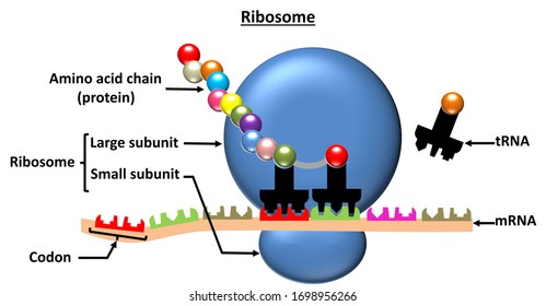
                  Ribosomes are small, tiny, spheroidal dense particles present in all living cells. The word ribosome is derived - 'ribo' from ribonucleic and 'somes' from the Greek word "soma" means 'body'. They are the sites of protein synthesis. These ribosomes are primarily found in both prokaryotic and eukaryotic cells. Ribosomes link amino acids together in the order that is specified by the messenger RNA (mRNA) and is of 150-200 Angstroms. A ribosome is made from complexes of RNAs and proteins, and is, therefore a ribonucleoprotein. Around 37-62% of ribosomes are made up of RNA and the rest is proteins. 

Structure of Ribosomes:

            Ribosomes Images, Stock Photos & Vectors | Shutterstock

                              The ribosomes are small particles present in large numbers in many cells. Each ribosome consists of two subunits:- a) larger subunit and
                                                                                  b)smaller subunit
a) The larger subunit bind to tRNA, amino acids, and the smaller subunit.
b)The smaller subunit binds to a larger subunit and mRNA pattern.
        Ribosomes are located in two regions of the cytoplasm. Some are found scattered in the cytoplasm and some are attached to the Endoplasmic reticulum. When the ribosomes are bound to ER, they are known as Rough endoplasmic reticulum. The bound and free ribosomes are similar in structure and are involved in protein synthesis. 
 Note:- The subunits of ribosomes are named according to their ability of sedimentation on a special gel called as "Svedberg" and hence svedberg units.
  • The prokaryotes have 70s ribosomes where each subunit consists of a small unit of 30s and a large subunit of 50s.
  • The eukaryotes have 80s ribosomes where each subunit consists of small unit of 40s  and a large subunit of 60s.
  • The ribosomes found in the chloroplasts or mitochondria of eukaryotes that consist of large and small units bound together with proteins into one 70s particle.
  • The number of ribosomes in a cell depends on the activity of the cell.
  • The ribosomes share a core structure and the two subunits fit together and work as one to translate the mRNA into a polypeptide chain during protein synthesis.
  • During protein synthesis, when multiple ribosomes are attached to the same mRNA strand, this structure is called as "polysome".
  • The location of the ribosomes in the cell determines what type of protein it makes.For eg; if the ribosomes are freely situated throughout the cell, it will make proteins that will be utilized within the cell itself.

                            A.2.1. Structure of the cell

Functions:-

                   The functions of the ribosomes can be explained in the following different ways:-
  1. The ribosomes are the major sites of protein synthesis.
  2. These ribosomes assemble amino acids to form specific proteins that are essential to carry out cellular activities.
  3. The genetic message from the mRNA is translated into the proteins during DNA translation.
  4. The ribosomes are also involved in DNA transcription, a process in which the proteins are produced; where deoxyribonucleic acid produces mRNA that is later converted into proteins.
  5. The mRNA is synthesized in the nucleus and is transported to the cytoplasm for further protein synthesis.
  6. The two subunits of ribosomes are bound around the polymers of mRNA in the cytoplasm; and proteins are then synthesized with the help of transfer RNA (tRNA).
Unit 7 - Protein Synthesis Diagram | Quizlet
protein synthesis diagrams


  • courtesy by google images                            


Thursday, 9 July 2020

Lysosomes

LYSOSOMES:

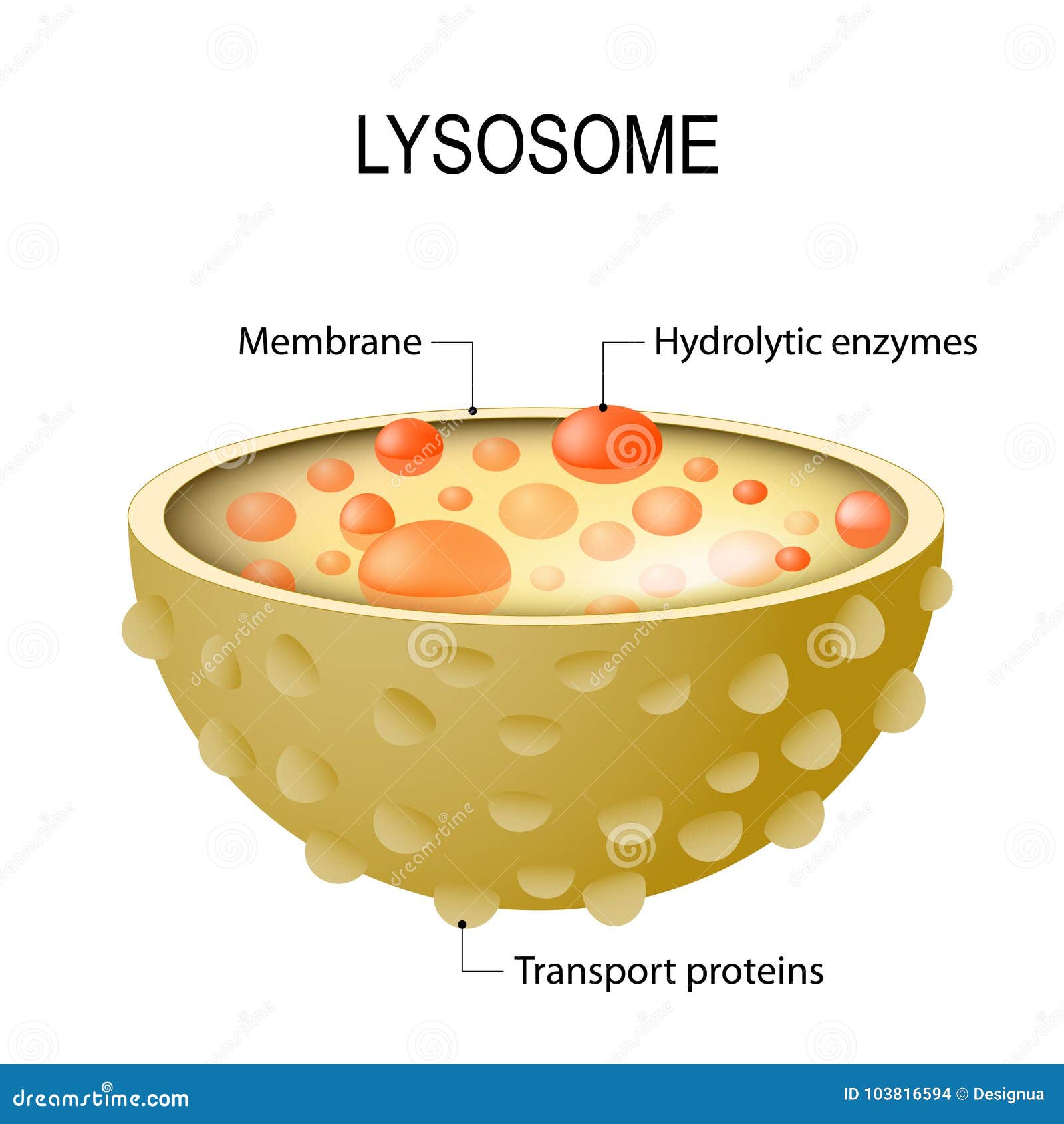
                             Lysosomes are membrane-bound tiny bags filled with the hydrolytic enzymes that are concerned with both intracellular and extracellular digestion. A typical lysosome is a lytic body that is capable of lysis. Lysosomes can occur freely in the cytoplasm. The word "lysosome" is made up of two words ie, 'lyso' means 'digestive' or 'breakdown', and 'soma' means 'body'. These lysosomes were first named as "pericanalicular bodies" because of their location but later it was renamed as lysosomes by"Christian de Duva" in the year 1955.
                           Lysosomes occur in most animal cells and in a few plant cells too, (in the form of vacuoles). They are most abundant in the cells which are related to enzymatic reactions such as liver cells, pancreatic cells, kidney cells, spleen cells, leukocytes, etc except in the RBCs.

                                      Lysosomes | Definition, Examples, Diagrams


Structure of lysosomes:

Anatomy Of The Lysosome. Vector Diagram For Medical Use Stock ...
                   Lysosomes are spherical dense bodies filled with large granules of digestive enzymes and acid phosphatases. Lysosomes are spherical in shape, but they are irregular in certain meristematic cells of roots. The size of the lysosomes usually ranges from 0.2-0.8 in diameter but may be exceptionally large as 8 microns in mammalian kidney cells and leucocytes. The lysosomes are bounded by a single-layered membrane in contrast to the double-layered membranes of other organelles. It is made up of proteins and lipids. 
            The interior of some lysosomes is uniformly solid while others have a very dense outer zone and a less dense inner zone. The inner of the lysosome is acidic with a pH of 4.8, but the pH of the surrounding cytosol is 7.2. The low pH is maintained by pumping the protons from the cytosol. Lysosomes are polymorphic structures because of their contents vary in the stages of digestion.


                                Cells Lysosome Transparent & PNG Clipart Free Download - YAWD


Types of Lysosomes:

  1. Primary lysosomes: These are small sac-like structures enclosing enzymes synthesized by the rough endoplasmic reticulum. They are also said to be storage granules as they store enzymes. The enzymes present in primary lysosomes are acid hydrolases.
  2. Secondary lysosomes: These are formed by the fusion of primary lysosomes with phagosomes. They contain engulfed materials and enzymes. 
  3. Residual lysosomes: The secondary lysosomes with the undigested wastes (residues) are called as residual lysosomes. The digested materials are diffused into the cell cytoplasm through the lysosomal membrane.
  Lysosomes mainly involve in these activities:

  1. Intracellular digestion:- It follows in various aspects:
  •   Autophagy: 
                            Autophagy refers to the lysosomal digestion of own cell components where 'auto' means 'self'' and 'phago' means 'eating'. Here the cell organelles, worn-out cells, dead cells, cell debris, and stored food materials are digested by the lysosomes. In autophagy, the organelle to be digested is enclosed by a membrane called 'isolation membrane' (derived from ER or Golgi body) 
The cell to be digested enters into this membrane and forms an "isolation body". This isolation body fuses with the lysosome to form autophagic vesicles and is digested. The digested particles now diffuse into the cytoplasm and are utilized by the cell for metabolic activities.


                                                                                 Lysosomal membrane traffi cking pathways. Lysosomes receive inputs ...

  • Heterophagy:
                               It is the lysosomal digestion of foreign materials. Here the cell digests the foreign or extracellular food materials. These food materials are taken into the cells by endocytosis such as phagocytosis.

  • Autolysis: 
                Autolysis refers to the killing of entire set of cells by breakdown of the lysosomal membrane. Here auto means 'self' and lysis means 'kill'. In autolysis, the lysosome digests its own cell; hence autolysis is called as "cellular autolysis". In this process, the lysosome ruptures inside its cell, and the released enzymes digest and degrade the cell. As the lysosome kills its own cell, it is called as "suicidal bag".Hence lysosomes are known as "Suicidal bags of cell"

2. Extracellular digestion:

                           The digestion of materials outside the cell is called "Extracellular digestion". On certain occasions, lysosomes release enzymes outside the cell by exocytosis and bring about digestion. For eg; this digestion occurs during bone erosion. The osteoclasts are rich in lysosomes. In the area of erosion, the lysosomes release enzymes outside the cell and bring about the extracellular digestion of bone.

Other functions of lysosomes are:-

  • Fertilization:- The acrosome of sperm ruptures and releases enzymes such as hyaluronidase, proteases, etc which dissolve the egg membrane and make a way for the entry of sperm into the egg.
  • Chromosomal breakage:-The lysosomes contain the enzyme deoxyribonuclease. This enzyme attacks the chromosome and brings about chromosomal breakages.
  • Programmed cell death is also caused by lysosomes.
  • Lysosomes also help in the developmental process.
  • Lysosomes function as a garage disposal system of the cell.
  • Autolysis occurs during amphibian metamorphosis, insect metamorphosis, menstruation, etc
  • Lysosomes bring about intracellular digestion and extracellular digestion.
                                                       
                            8 Main Functions of Lysosomes | Biology
  • courtesy by google images


Monday, 15 June 2020

Chloroplasts


CHLOROPLASTS:
                                         Chloroplasts are membrane-bound plastids that contain a network of membranes embedded in the liquid. These are the organelles that conduct photosynthesis with the help of pigment called as "Chlorophyll". It is this pigment that imparts a green color to the leaves and other parts of the plants and captures light energy from the sunlight. Chloroplasts are important because if there were no chloroplasts, plants cannot produce energy in the daylight and the whole survival of living organisms become difficult. These are present only in plant cells and are absent in animal cells. These chloroplasts are also found in bacteria, blue-green algae, etc. These are the sites of photosynthesis.
               The word chloroplast is derived from the Greek words 'chloros' meaning "green" and 'plastes 'meaning "formed". Chloroplasts were first discovered in the early 17th century, by "Antony Van Leeuwenhoek" and "Nehemiah Grew". These chloroplasts can be found in the mesophyll cells of plants where are they usually 30-40 per mesophyll cells. Chloroplasts are a type of plastids, some other types are leucoplasts, chromoplasts, etc.

                                    File:Figure 08 01 05.png - Wikimedia Commons

          Chloroplasts are the part of the plant and algal cells that carry out photosynthesis, the process of converting light energy into energy stored in the form of sugar and other organic molecules. The process of photosynthesis has two stages. In the first stage, light-dependent reactions occur which captures sunlight through chlorophyll to form Adenosine triphosphate (ATP), Nicotinamide adenine dinucleotide phosphate (NADPH). In the second stage, light-independent reactions occur (also known as Calvin cycle), where the electrons carried by NADPH convert inorganic carbon dioxide to an organic compound called as carbohydrate. This is known as CO2 fixation. Carbohydrates and other organic molecules can be stored and used for energy. 
Structure of chloroplast:
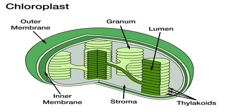
                     Chloroplasts are oval-shaped and consist of two membranes, ie, the outer membrane that forms the external surface of it and an inner membrane that lies just beneath it. An intermembrane is present between the outer and inner membrane. The space within the inner membrane is called "stroma". It also contains many small disc-shaped sacs called "thylakoids" that are stacked upon one another. The main components of the chloroplast are:
1.     Envelope: 
a)   Outer membrane: The entire chloroplast is bounded by an outer double unit membrane. It is a semi-porous membrane that is permeable to small molecules and ions that diffuse easily. It is not permeable to large molecules or proteins.
b)   Inner membrane: It is present just beneath the outer membrane and forms a boundary to the stroma. Fatty acids, lipids, carotenoids, etc, are synthesized in this membrane. It also regulates the passage of materials in and out of the membrane.
c)  Intermembrane space: The space between the inner and outer membrane is called intermembrane space. It is usually thin and about 10-20 nanometres.


       Atp - Lessons - Tes Teach

                                                 
    2. Stroma:
  • Stroma is an alkaline, aqueous fluid that is present in the inner membrane of the chloroplast.
  • It is protein-rich and contains about 50% of the proteins of the chloroplast.
  • Stroma also contains chromosomes and DNA molecules.
  • It is the place where carbon dioxide fixation takes place and the synthesis of sugar, starch, and other proteins takes place.
  • It is a gel-like fluid that surrounds the thylakoids.
    3. Thylakoids:
  • A disc of flattened membranous sacs is called as Thylakoids and a collection of these are called a Thylakoid system.
  • Chlorophyll is found in the thylakoids and is the site of light reaction and photosynthesis processes.
  • These thylakoids are arranged in stacks called "grana". Each granum consists of 10-30 thylakoids.
  • The number of thylakoids per granum varies from 1-50 or more. For eg; there may be a single thylakoid in red algae, paired algae in chrysophytes and triple and multiple in green algae and higher plants.
  • Structural lipids of thylakoids are present here that include glycolipids, sulpholipids, and a few phospholipids, etc.
  • These structural lipids are highly unsaturated which counter a high degree of humidity to the membrane of thylakoids.
  • The protein components of thylakoids involve in the following complexes such as Photosystem-I, Photosystem-II, Cytochrome, ATP synthesis, Light-harvesting compound.


                                   NCERT Class XI Biology: Chapter 13 - Photosynthesis In Higher ...


Functions of Chloroplasts:
  • Chloroplasts are the site of photosynthesis that comprises of light-dependent and light-independent reactions to convert solar energy into usable and stored energy.
  • These involve in the regulatory functions and helps in the synthesis of proteins and lipids.
  • Production of ATP and NADPH by phosphorylation.
  • Light reactions take place in membranes of thylakoids
  • Dark reactions take place in the stroma.
  • Conversion of PGA into sugars and starch that can be stored.


  • Courtesy by google images